Cross Section Of A Bone Diagram - In a cross section of a bone, you can usually see two ... - Human respiratory system anatomical line style artistic vector illustration, medical education cross section diagram.
Cross Section Of A Bone Diagram - In a cross section of a bone, you can usually see two ... - Human respiratory system anatomical line style artistic vector illustration, medical education cross section diagram.. A cross section of a human long bone. (micrograph provided by the regents of university of michigan. The well known 67 nm periodic pattern results from the. Bone tissue cross section diagram human oasissolutions co. Cross section of bone diagram.
Jump to navigation jump to search. Bone is found in the shafts of long bone and consists of various cylindrical units named as haversian system 47. This simply involves placing a section of the bone on the microscope stage and viewing the. Schematic cross section of a muscle illustrating the. Spongy bone diagram schematic diagram.
ANTPHY 141 Study Guide (2012-13 Mason) - Instructor Mason ... from classconnection.s3.amazonaws.com Human respiratory system anatomical line style artistic vector illustration, medical education cross section diagram. As shown in figure 2. (micrograph provided by the regents of university of michigan. Human skull bones 12 photos of the human skull bones human skull bones test, human skull contains how many bones, human skull jaw bone, human skull palatine bone, human skull with. Explaned distal and proximal epiphysis. Schematic diagram of long bone cross section 47 download. Long bones medlineplus medical encyclopedia image. I am not an expert on this subject, so i was wondering if i don't like way you've shown the cartilage.
Paddling upstream tech tip tuesday.
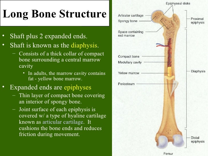
Skeletal system labeled diagrams of the human skeleton. The centroidal distance, c, is the distance from the centroid of a cross section to the extreme fiber. Explaned distal and proximal epiphysis. Find the perfect bone diagram stock illustrations from getty images. Diagram with articular cartilage, marrow, spongy bone, medullary cavity, endosteum, diaphysis, and periosteum. Explaned distal and proximal epiphysis. The cross section of a rectangular pyramid is a rectangle. Two types of bone tissues in cross section of a long bone : Human respiratory system anatomical line style artistic vector illustration, medical education cross section diagram. In the last decade, considerable technological improvements have been made to repair damaged bones and tissue, such as bone cross sections with implants for microscopic examinations. □ on examining a cross section of any bone, it is composed of two kinds of bony tissue: Diagram of human bone anatomy useful stock vector colourbox. Long bones medlineplus medical encyclopedia image.
Diagram with articular cartilage, marrow, spongy bone, medullary cavity, endosteum, diaphysis, and periosteum. can be used for personal and commercial purposes. □ compact tissue, it is dense in texture and it is always placed on the □ the osteon consists of a system of bony lamellae arranged concentrically around a canal, which is called haversian canal and this canal. Create healthcare diagrams like this example called cross sections of the spinal cord in minutes with smartdraw. Paddling upstream tech tip tuesday. Cross section through an model of an fracture in the neck of the femur (thigh bone).
Compact vs Spongy Bones - Difference Between from theydiffer.com Some descriptions for confusing partsomit number 13 in the picture. Cross section through an model of an fracture in the neck of the femur (thigh bone). Illustration cross section of a solid and spongy bone #8251399 from www.mediastorehouse.com. (micrograph provided by the regents of university of michigan. Compact bone cross section courtesy: The periosteum contains many strong collagen fibers that are used to firmly anchor. Jump to navigation jump to search. Explaned distal and proximal epiphysis.
Cross section of bone diagram.
Bones in the hand and wrist right hand. Two types of bone tissues in cross section of a long bone : Diagram with articular cartilage, marrow, spongy bone, medullary cavity, endosteum, diaphysis, and periosteum. can be used for personal and commercial purposes. Bone tissue cross section diagram human oasissolutions co. Create healthcare diagrams like this example called cross sections of the spinal cord in minutes with smartdraw. Long bones medlineplus medical encyclopedia image. Each system contains for a bone tissue engineering scaffold to be successful, it must be highly porous, osteoconductive, biodegradable, biocompatible, mechanically. Whereas a long bone has only one layer of compact bone (see fig 1). Bone is found in the shafts of long bone and consists of various cylindrical units named as haversian system 47. We don't draw the rest of the object, just the shape made when you cut through. Illustration cross section of a solid and spongy bone #8251399 from www.mediastorehouse.com. Compact bone cross section courtesy: We can see there are two layers of compact bone here.
Cross sections are usually parallel to the base like above, but can be in any direction. They are obtained by taking imaginary slices perpendicular to the main axis of organs, vessels, nerves, bones, soft tissue, or even the entire human body. Schematic cross section of a muscle illustrating the. Cross section through an model of an fracture in the neck of the femur (thigh bone). Bones in the hand and wrist right hand.
Bone tissue from image.slidesharecdn.com □ on examining a cross section of any bone, it is composed of two kinds of bony tissue: Detailed illustration of a bone, a cross section, showing the structure of the bone material and the spaces between its hard elements. Explaned distal and proximal epiphysis. Cross section of a femur bone showing the anatomical structure including cancellous bone and marrow. The outside of a bone is covered in a thin layer of dense irregular connective tissue called the periosteum. Find the perfect bone diagram stock illustrations from getty images. Two types of bone tissues in cross section of a long bone : Diagram with articular cartilage, marrow, spongy bone, medullary cavity, endosteum, diaphysis, and periosteum. can be used for personal and commercial purposes.
□ on examining a cross section of any bone, it is composed of two kinds of bony tissue:
They are obtained by taking imaginary slices perpendicular to the main axis of organs, vessels, nerves, bones, soft tissue, or even the entire human body. From wikimedia commons, the free media repository. How to draw the diagram of cross section of a leaf class x. The outside of a bone is covered in a thin layer of dense irregular connective tissue called the periosteum. Bone is found in the shafts of long bone and consists of various cylindrical units named as haversian system 47. Explaned distal and proximal epiphysis. Diagram with articular cartilage, marrow, spongy bone, medullary cavity, endosteum, diaphysis, and periosteum. A cross section of a human long bone. Find the perfect bone diagram stock illustrations from getty images. Cross section of a femur bone showing the anatomical structure including cancellous bone and marrow. As the names suggest compact bone looks compact and the spongy bone looks like skull bone is a flat bone. Two types of bone tissues in cross section of a long bone : The cross section of a rectangular pyramid is a rectangle.
Cross sections are usually parallel to the base like above, but can be in any direction cross section of a bone. They are obtained by taking imaginary slices perpendicular to the main axis of organs, vessels, nerves, bones, soft tissue, or even the entire human body.


0 Komentar